Particle Image Analyzer
- Products
- Petroleum & Oil Testing
- Tablet Drug Tester
- Refrigeration & Cryogenic
- Life Sciences
- Laboratory
- Spectrometer
- Spray Dryer
- Rotary Evaporator
- Reactor
- Sterilizing & Cleaning
- Water Quality Analyzer
- Chemical Analysis
- Physical Testing
- Centrifuge
- Pathology Equipment
- Optical
- Ultrasonic Homogenizer
- Packaging Testers
- NDT
- Agriculture & Food
- Hazardous Chemical Detection
- Fusion Machine
- Testing Chamber
- Filter Integrity Tester
- Industry Testing Device
- Featured
More info
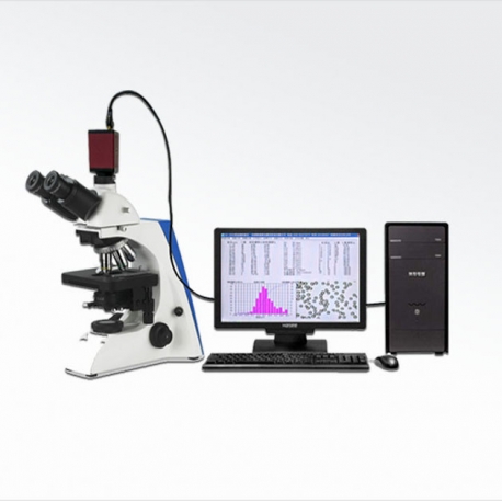
PA-2000AI image analysis system consists of optical microscope, digital CCD camera, image processing and analysis software, computer, printer and other parts. Because the powders have different shapes, when testing with a laser particle size analyzer, the PA-2000AI image analyzer can be used to more accurately visualize the particle size and shape and control the product quality.
Particle image test principle:
Particles are imaged on an imager. The smallest unit that makes up an image is pixels, and each pixel has a specific size. By counting the number of particles and the number of pixels contained in each particle, the diameter of the equal area circle and the aspect ratio of each particle are calculated, and then all particles are counted to obtain information such as particle size distribution.
The main process of image analysis is:
The sample is enlarged and imaged by the microscope, and the clear particle morphology image taken by the camera is transmitted to the computer. The morphology of the powder particles is directly observed on the computer. The number, area, perimeter, diameter, and volume of the particles are calculated and analyzed by proprietary software. , Long-diameter ratio, short-diameter ratio, roundness coefficient and other distribution data and images. Data distribution charts such as D10, D50, D90, D97, average particle size, and surface area are also provided.
Application area
Diamond, silicon carbide, wollastonite, quartz powder, barium sulfate, graphite, lithium cobaltate, boron carbide, white corundum, cerium oxide mica powder, carbon powder, metal powder and other mineral powders:
Various powders or slurries such as glass beads, cement, medicine, food, pigment, dust, pesticide, catalyst, etc.
It can also be used to verify other means and methods of granularity testing.
Technical Parameters
Model | PA-2000AI |
Analysis Project | Particle size distribution, aspect ratio distribution, circularity distribution, etc. |
Test Range | 0.5 ~ 3000μm |
Maximum magnification | 4000 times |
Maximum resolution | 0.1μm |
microscope | Transmission microscope (optional domestic / metallographic / import) |
Digital Camera (CCD) | 3MP / 5MP |
eyepiece | 10X, 16X |
Objective lens | 4X, 10X, 20X, 40X, 100X |
Ruler scale | 10μm |
Interface method | USB interface for high-speed and stable transmission |
Report output
After processing the image, the computer automatically counts the number of particles, area, circumference, volume, diameter, circularity distribution, aspect ratio distribution, particle size distribution data, and D10, D50, D90, D97, average Characteristic values such as particle size and surface area.
The color thumbnail of the sample can be displayed in the report, and multiple information such as the sample name, test unit, and dispersion medium can be entered into the report header. Multiple report formats can be selected.


